Mechanism of Dysfunction
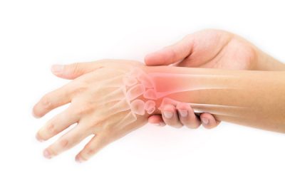
The predisposing factors for the development of this condition began with a misalignment dysfunction at the level of the distal radial and ulna casing the first row of the wrist to compensates and becomes hypermobile which overtime causes the soft tissue ligamentous and muscle support to become overloaded and compromised allowing the biomechanical stress to reach the level of the cartilage and initiating the degenerative joint disease.
Is important to note that for the cartilage to become injured, the previous defence mechanisms have to have failed to allow the biomechanical stress to damage the cartilage, therefore, the treatment care must aim to restore the health of the entire ankle/foot protective structures.
Assessment Protocol
The entire upper extremity biomechanical chain must be evaluated as part of the foot/Ankle analyses as per the neurological and mechanical influences of the spine, shoulder, and elbow.
Clinical assessment to identify the key joint dysfunctions of the wrist that have contributed to this condition. Soft tissue analysis to pinpoint the level of irritation in the soft tissue support.
X-ray
Anterior – Posterior (AP) X-ray wrist view is essential for proper diagnosing the master joint of the wrist (Radial – Ulna) and the alignment of the scaphoid and triquetrum.
Lateral Xray view is important to check the degree of the total arch compromised and the direction of misalignment of the lunate bone
MRI
Wrist/hand MRI is essential for visualizing the extent of injury on the muscle/tendon ligamentous and cartilage layers.
Locate the exact injury point; Allows the treatment to be more specific during the application of the treatment modalities.
Identify the extent of tissue damage and the presence of scar tissue; Provides valuable information regarding prognosis and the application of friction soft tissue modalities to aid on scar tissue removal.
Rule out stress fractures not visible by the x-ray.
Treatment protocol
The treatment care should aim to restore the wrist and hand alignment, the soft tissue muscles/tendon and ligamentous health as well as stimulating and remodelling the cartilage growth.
Specific adjustments of key bones of the wrist followed by a rehabilitation regime to strengthen the entire soft tissue support of the wrist and hand.
Soft tissue and cartilage healing protocol
Application of Low-level Laser and PEMF directly over the injured tissues to aid on the cellular level of healing as well as improving the microcirculation for the area.
Friction soft tissue therapy helps to reduce dysfunctional scar tissue
Specific stretches and strengthening to improve the resilient of the soft tissue support
Specific selected essential oil application to enhance healing
Dry Needling to promotes blood flow and enhance the soft tissue and cartilage healing.
Depending on the level injury and chronicity, a minimum of 6 weeks up to 12 weeks of treatment care may be necessary to resolve this condition.


