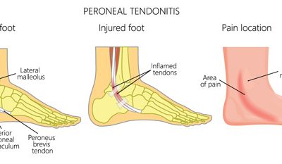
Mechanism of Dysfunction
There is a typical mechanical injury pattern that contribute to this condition. Generally, the patient is in a weight bearing position with the foot slightly plantar flexed (pointed down) and rotated outward (everted), the increased mechanical torsion with the foot held in that position, predisposed to this sprain injury.
Is important to note that for the ligament to become injured, the previous defence mechanisms have to have failed to allow the biomechanical stress to damage the ligament, therefore, the treatment care must aim to restore the health of the entire ankle/foot protective structures.
Assessment Protocol
The entire lower extremity biomechanical chain must be evaluated as part of the foot/Ankle analyses as per the neurological and mechanical influences of the spine, hip, and knee.
Clinical assessment to identify the key joint dysfunctions of the Ankle/Foot (Talus, Fibula, Navicular, and calcaneus) that have contributed this condition. Soft tissue analysis to pinpoint the level of irritation in the ligaments and muscles/tendons.
X-ray analysis
Anterior – Posterior (AP) X-ray ankle view is essential for proper diagnosing the master joint of the ankle (Tibia-Talus) as the origin of the eversion sprain begun with a dysfunctional of this joint.
Lateral Xray view is important to check the further misalignment of the talus and the possible involvement of navicular and calcaneus.
MRI
Ankle/foot MRI is essential for visualizing the extent of injury on the muscle/tendon and ligamentous layers.
Locate the exact injury point; Allows the treatment to be more specific during the application of the treatment modalities.
Identify the extent of tissue damage and the presence of scar tissue; Provides valuable information regarding prognosis and the application of friction soft tissue modalities to aid on scar tissue removal.
Rule out stress fractures not visible by the x-ray.
Treatment protocol
The treatment care should aim to restore the foot/ankle alignment and the soft tissue muscles/tendon and ligamentous layers.
Specific adjustments of key bones of the ankle and foot (Talus, Fibula, Navicular, Calcaneus and Cuboid) followed by a rehabilitation regime to strengthen the entire soft tissue support of the ankle.
Application of Low-level Laser and PEMF directly over the injured tissues to aid on the cellular level of healing as well as improving the microcirculation for the area.
Friction soft tissue therapy helps to reduce dysfunctional scar tissue
Specific stretches and strengthening to improve the resilient of the soft tissue support
Functional tape might be used to reposition the talus and fibula
Specific selected essential oil application to enhance healing
Depending on the level injury and chronicity, a minimum of 6 weeks up to 12 weeks of treatment care may be necessary to resolve this condition.
Related Conditions
Peroneal tendinitis
Mechanism of...
Tibialis anterior tendinitis
Tibialis...
Ankle and foot fractures
Mechanism...


